Drlogy
Healthcare organization
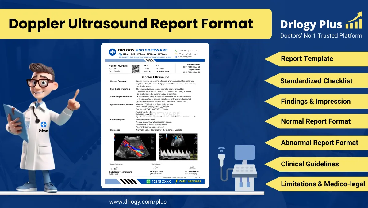
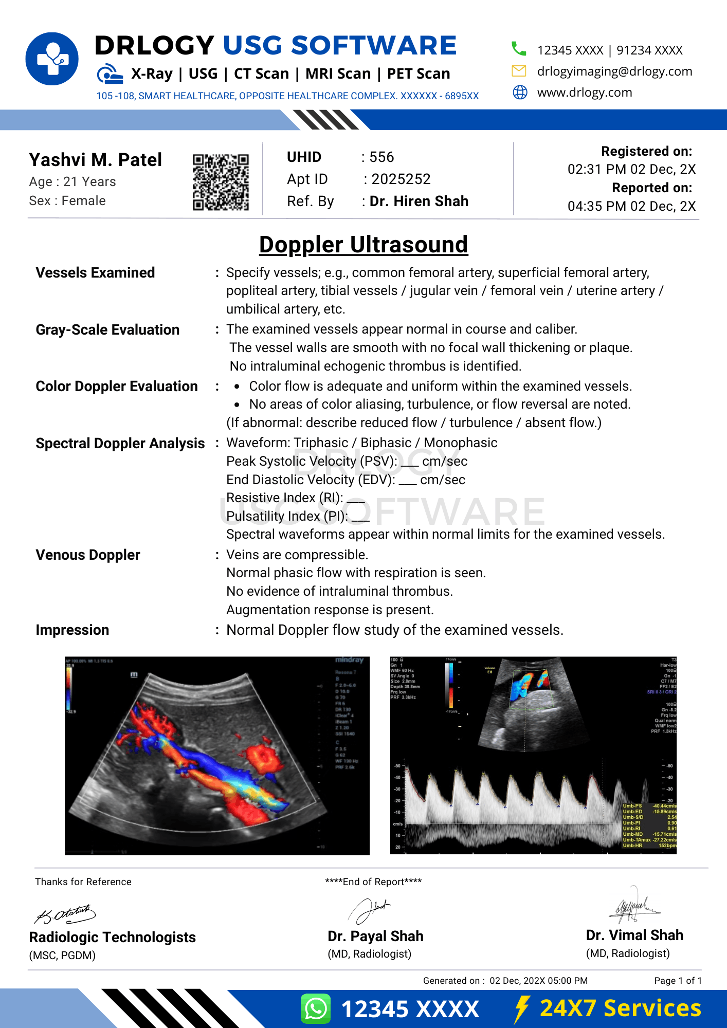
Doppler Ultrasound Report Format for Radiologists
What Is a Doppler Ultrasound Report Format?
A Doppler ultrasound report format is a standardized professional structure for documenting blood flow assessment using spectral, color, and power Doppler techniques with precise vascular terminology.
It functions as a formal clinical communication record supporting diagnosis, referrals, follow-up, treatment planning, and longitudinal vascular comparison.
It is a medico-legal document defining examination scope, technical adequacy, objective hemodynamic measurements, conservative interpretation, and documented limitations.
Check:
Best AI-Based Ultrasound Reporting Software for Radiology Center
Clinical Importance of a Standardized Doppler Ultrasound Report Format
- Diagnostic clarity through structured documentation of vessel patency, flowection, velocity parameters, and waveform morphology.
- Inter-doctor communication using uniform hemodynamic terminology understood by radiologists, clinicians, surgeons, and interventional specialists.
- Reporting consistency across different vascular beds, serial examinations, and multi-radiologist environments.
- Patient safety by reducing omission of critical parameters such as peak systolic velocity, resistive index, and flow symmetry.
- Medico-legal protection through objective measurements, conservative impressions, and clearly stated technical limitations.
A standardized Doppler ultrasound report format ensures accuracy, reproducibility, and audit-ready vascular documentation.
Why Manual Reporting Often Fails to Maintain Standardization at Scale
- Inter-radiologist variability in velocity measurement reporting, waveform interpretation, and terminology usage.
- Missed sections in high-volume settings such as incomplete documentation of bilateral comparison or absent spectral analysis.
- Terminology inconsistency including vague terms like reduced flow without quantitative support.
- Audit challenges due to narrative reports lacking structured hemodynamic data fields.
Structured reporting improves reliability and medico-legal robustness while preserving clinical judgment.
Indications for Doppler Ultrasound
- Evaluation of arterial or venous patency
- Assessment of suspected vascular stenosis or occlusion
- Venous thrombosis evaluation
- Peripheral arterial disease assessment
- Organ-specific vascular evaluation
- Follow-up of known vascular abnormalities
Doppler ultrasound indications guide protocol selection and parameter emphasis.
Pre-Examination Details to Be Documented
- Patiententifiers including name, age, unique, accession number, study date, and time.
- Referral details including referring clinician and specific vascular query.
- Clinical notes including symptoms, duration, relevant medical history, and prior vascular imaging if provided.
- Preparation status including fasting or positioning requirements where applicable.
- Safety checks including correct patient verification and side confirmation.
How Reporting Software Ensures Complete Pre-Examination Documentation
- Mandatory field enforcement for indication, vascular territory, and laterality.
- Safety checklist compliance ensuring correct patient and vesselentification.
- Clinical note traceability linking referral information with Doppler findings.
- Implementation example: Drlogy Radiology Reporting Software offers structured Doppler templates with compulsory velocity and waveform documentation fields.
Standard Sections of a Doppler Ultrasound Report Format
- Patient & Study Information
- Clinical History / Indication
- Technique / Protocol
- Findings (vessel-wise hemodynamic assessment)
- Impression / Conclusion
- Limitations of the Study
- Recommendations & Follow-Up (if applicable)
Patient & Study Information Section
This section establishes accountability and traceability:
- Patient demographics andentifiers
- Study date, time, and accession number
- Referring clinician details
- Doppler examination type and vascular territory
- Comparison with prior Doppler studies when available
Clinical History / Indication Section
- Indication for Doppler evaluation
- Symptom description and laterality
- Relevant medical or surgical history
- Prior imaging or intervention details if provided
Documentation must remain concise and clinically relevant.
Technique / Protocol Section
- Positioning: supine, prone, or limb positioning depending on vascular territory.
- Approach: high-frequency linear or curvilinear transducer selection.
- Doppler modes: color Doppler, power Doppler, and spectral Doppler usage documented.
- Angle correction: standardized insonation angle documentation.
- Measurements: PSV, EDV, RI, PI as applicable.
Technique documentation defines measurement validity and interpretive confidence.
Findings Section – Organ/System-Wise Reporting
Vessel Assessment
- Vesselentification and laterality
- Patency and luminal characteristics
- Flowection and symmetry
- Spectral waveform morphology
Velocity Measurements
- Peak systolic velocity
- End-diastolic velocity
- Calculated indices where applicable
Comparative Evaluation
- Side-to-side comparison
- Proximal and distal flow assessment
Objective description must precede interpretive statements.
Impression / Conclusion Section
- Concise summary of hemodynamic findings
- Conservative, non-definitive language
- Avoidance of therapeutic recommendations
- Correlation with clinical findings advised
Limitations of the Study
- Suboptimal insonation angle
- Patient body habitus
- Vessel depth or calcification
- Motion artifacts
Documenting limitations is essential for medico-legal transparency.
Recommendations & Follow-Up (If Applicable)
- Clinical correlation with symptoms
- Follow-up Doppler evaluation when indicated
- Additional imaging only when clinically appropriate
Recommendations must remain conservative and protocol-aligned.
Normal Doppler Ultrasound Report Format (Sample)
- Patient & Study Information:
- Patient: [Name], [Age]
- Study Date: [DD-MM-YYYY]
Examination: Doppler Ultrasound
Clinical History / Indication:
Vascular evaluation.
Technique / Protocol:
Color and spectral Doppler evaluation performed.
Findings:
The examined vessels show normal patency with laminar flow. Spectral waveforms and velocity parameters are within expected limits.
Impression / Conclusion:
No significant Doppler abnormality detected.
Limitations:
No significant technical limitation noted.
Abnormal Doppler Ultrasound Report Format (Sample)
- Patient & Study Information:
- Patient: [Name], [Age]
- Study Date: [DD-MM-YYYY]
Examination: Doppler Ultrasound
Clinical History / Indication:
Suspected vascular pathology.
Technique / Protocol:
Doppler evaluation performed.
Findings:
Altered flow velocities and waveform changes are noted in the examined vessel as described.
Impression / Conclusion:
Doppler findings as described. Clinical correlation is advised.
Limitations:
Assessment limited by vessel depth.
How Drlogy Radiology Reporting Software Standardizes These Report Formats
- Template-driven reporting ensuring complete Doppler parameter documentation
- Impression safety controls enforcing conservative language
- Uniform formatting across vascular territories
- AI-enabled reporting assistance under radiologist supervision
- Audit-ready documentation supporting quality assurance
10 Key Clinical Guidelines for an Effective Doppler Ultrasound Report Format
- Identify vessels clearly.
- Document laterality consistently.
- Use standardized Doppler angles.
- Record velocity measurements accurately.
- Describe waveform morphology objectively.
- Compare bilateral findings.
- Separate findings from impression.
- Use conservative terminology.
- Document technical limitations.
- Maintain consistent report structure.
Adherence improves diagnostic reliability and medico-legal safety.
Common Reporting Errors to Avoid
- Omission of velocity values
- Vague flow description without quantification
- Incomplete bilateral comparison
- Overinterpretation of isolated findings
- Missing limitation statements
Avoiding these errors strengthens report credibility.
Medico-Legal Considerations in Radiology Reporting
- Objective measurement documentation
- Standardized terminology usage
- Conservative impression language
- Explicit limitation statements
- Clear accountability
- Audit readiness
- Appropriate disclaimers
Structured Reporting vs Narrative Reporting
| Aspect | Structured | Narrative |
|---|---|---|
| Completeness | Protocol-driven | Variable |
| Consistency | High | Operator dependent |
| Audit readiness | Strong | Limited |
| Efficiency | Optimized | Variable |
| Legal safety | Enhanced | Variable |
Role of Technology in Radiology Reporting
- PACS and RIS integration
- Voice dictation with templates
- AI-assisted formatting
- RIS-based structured Doppler templates
- Vascular workflow tools
Technology enhances consistency without replacing professional judgment.
Why High-Volume Radiology Centers Prefer Software-Based Reporting Formats
- Faster turnaround time
- Improved quality assurance
- Multi-radiologist consistency
- Scalability across vascular studies
- Reduced omission errors
- Standardized hemodynamic documentation
- Enhanced medico-legal protection
Frequently Asked Questions (FAQs)
What defines a standard Doppler ultrasound report format?
A structured format documenting vessel patency, flow characteristics, velocity parameters, impression, and limitations using standardized terminology.
Is quantification mandatory in Doppler reporting?
Quantitative parameters are recommended to support objective interpretation.
How should equivocal Doppler findings be reported?
Using objective measurements with conservative impression and clinical correlation advice.
Why are limitations essential in Doppler ultrasound reports?
They document technical constraints and support medico-legal defensibility.
Key Takeaways for Radiology Professionals
- Use standardized Doppler reporting structure consistently.
- Document velocities and waveforms objectively.
- Maintain conservative impression language.
- Explicitly state technical limitations.
Structured Doppler reporting improves vascular assessment accuracy and medico-legal robustness.
Expert Picks
Final Conclusion
A standardized Doppler ultrasound report format is essential for accurate vascular communication, reliable follow-up, and medico-legal safety in diagnostic imaging practice.
Structured reporting software supports consistency, completeness, and conservative interpretation while aligning with real-world radiology workflows and established professional standards.