Drlogy
Healthcare organization
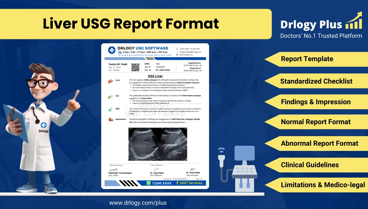
Liver USG Report Format for Radiologists
What Is a Liver USG Report Format?
A Liver USG report format is a standardized structure for documenting liver ultrasonography findings with consistent terminology, measurements, and organ-wise logic.
It supports diagnostic interpretation, referral decisions, follow-up planning, and interval comparison across serial studies in routine and high-volume reporting environments.
It is a medico-legal record defining examination scope, technique, observations, impression wording, and limitations, aligned with accepted radiology reporting standards.
Check:
Explore the Best AI-Based Ultrasound Reporting Software for Radiologists
Clinical Importance of a Standardized Liver USG Report Format
- Diagnostic clarity by ensuring uniform documentation of liver size, echotexture, surface contour, focal lesions, and vascular/biliary findings.
- Inter-doctor communication by using consistent descriptors that are interpretable across hepatology, gastroenterology, surgery, oncology, and primary care teams.
- Reporting consistency by reducing inter-radiologist variation in lesion description, vascular measurements, and impression phrasing.
- Patient safety by decreasing omission errors for key observations (e.g., portal vein caliber/flow, intrahepatic biliary dilatation, ascites).
- Medico-legal protection by documenting technique, limitations, and conservative impressions that remain defensible during audit or dispute review.
A standardized format improves reliability of liver ultrasound reporting across different operators, machines, and follow-up timepoints.
Why Manual Reporting Often Fails to Maintain Standardization at Scale
- Inter-radiologist variability in liver echogenicity terminology, lesion descriptors, and impression reduces reproducibility.
- Missed sections in high-volume settings commonly affect vascular assessment, biliary comments, and documentation of limitations.
- Terminology inconsistency (e.g., heterogeneous vs coarse echotexture vs altered echotexture) complicates clinician interpretation and follow-up comparisons.
- Audit challenges arise because free-text reports are difficult to benchmark, search, and quality-check against standard reporting requirements.
Software-assisted structured reporting improves completeness and uniformity while preserving radiologist judgment and interpretive autonomy.
Indications for Liver USG
- Abnormal liver function tests or biochemical cholestasis patterns
- Suspected fatty liver disease or chronic liver disease evaluation
- Hepatomegaly or suspected splenomegaly evaluation in clinical examination
- Right upper quadrant pain with suspected hepatobiliary origin
- Known hepatitis, cirrhosis, or portal hypertension follow-up
- Screening or follow-up for focal liver lesions in known malignancy or chronic liver disease
- Assessment of jaundice with suspected biliary obstruction (as part of hepatobiliary evaluation)
- Ascites evaluation with assessment for liver morphology and portal venous system features
- Post-intervention follow-up (post-biopsy, post-ablation, post-surgical evaluation) as clinically indicated
A concise indication improves exam targeting and supports clinically relevant impression formulation.
Pre-Examination Details to Be Documented
- Patiententifiers: full name, age, sex, unique, accession number, and study date/time.
- Referral details: referring clinician, department/specialty, and clinical question to be answered.
- Clinical notes: relevant history such as hepatitis status, alcohol-related risk, metabolic risk factors, known malignancy, prior liver lesions, and prior imaging summary where available.
- Preparation status: fasting status and adequacy, and any factors affecting acoustic window.
- Safety checks: confirmation of correct patient/study, documentation adequacy, and procedural notes where relevant (e.g., immediate post-procedure scan context).
Complete pre-examination documentation strengthens interpretive accuracy, follow-up comparability, and medico-legal defensibility.
How Reporting Software Ensures Complete Pre-Examination Documentation
- Mandatory field enforcement ensures criticalentifiers, referral details, and indication are completed before a report can be finalized.
- Safety checklist compliance supports standardized documentation steps such as correct patient verification, study labeling, and preparation status capture.
- Clinical note traceability links indication, relevant history, and prior study references within the reporting workflow for longitudinal comparison.
- Structured field prompts reduce omission of key liver-specific elements (size, echotexture, focal lesion characterization, portal vein assessment).
- Implementation example: Drlogy Radiology Reporting Software can be configured to enforce these fields and maintain consistent liver USG templates across users.
Documentation controls improve reporting completeness without changing clinical responsibility or interpretive authority.
Standard Sections of a Liver USG Report Format
Universally accepted sections for a professional liver USG report include:
- Patient & Study Information
- Clinical History / Indication
- Technique / Protocol
- Findings (structured liver assessment with relevant adjacent systems when applicable)
- Impression / Conclusion
- Limitations of the Study
- Recommendations & Follow-Up (if applicable)
A consistent section order improves readability, audit readiness, and rapid clinical interpretation.
Patient & Study Information Section
A robust metadata section should include:
- Patient name, age, sex, and uniqueentification number
- Study date and time, facility/unit, and accession/study number
- Referring clinician details and referral source
- Examination name: Liver USG (with Doppler details if performed)
- Prior studies reference (date and modality) when relevant and available
Clear metadata supports correct clinical linkage, follow-up, and medico-legal traceability.
Clinical History / Indication Section
Concise, clinically relevant documentation should include:
- Primary clinical concern (e.g., abnormal LFTs, suspected CLD, lesion follow-up)
- Known liver disease history (viral hepatitis, chronic liver disease, cirrhosis, portal hypertension)
- Relevant oncology history if lesion evaluation is requested
- Current symptoms or signs relevant to hepatobiliary pathology when provided
- Prior imaging summary (only key facts) and comparison request clarity
Avoid speculative statements; document only provided or verified clinical context.
Technique / Protocol Section
Technique documentation should reflect real-world ultrasound practice and should include:
- Positioning: supine, left lateral decubitus, intercostal approach as required for acoustic windows.
- Equipment: transducer type (curvilinear) and frequency range appropriate for body habitus; linear probe use for superficial lesions when indicated.
- Protocol scope: focused liver evaluation with assessment of hepatic parenchyma, liver surface, focal lesions, intrahepatic ducts, porta hepatis, and relevant vessels.
- Doppler usage: documentation of color/spectral Doppler assessment if performed, including portal vein and hepatic veins when clinically relevant.
- Breathing maneuvers: breath-hold or deep inspiration used for subcostal/intercostal views where needed.
- Preparation: fasting status noted; limitations if fasting inadequate or bowel gas interferes.
Technique documentation clarifies scope and supports defensible interpretation, especially when limitations exist.
Findings Section – Organ/System-Wise Reporting
A liver USG findings section should be structured, objective, and reproducible. Best practices include:
- Objective description first: describe what is seen, then interpret conservatively in the impression.
- Normal vs abnormal explicitly stated: normal structures should still be documented to confirm assessment completeness.
- Measurements and localization: liver size measurement method documented; lesion size in three dimensions where possible; segmental location when feasible; relation to capsule/porta hepatis.
- Standardized descriptors: echogenicity, echotexture, margins, posterior acoustic features, vascularity pattern (if Doppler), and ancillary signs.
- Relevant associated structures: intrahepatic ducts, common hepatic duct/CBD comment if part of the exam request; portal venous system and hepatic veins.
- Comparison: where prior imaging exists, include interval change phrasing cautiously (e.g., “appears similar,” “no significant interval change seen,” “interval increase cannot be excluded” when technique differs).
Liver Size and Morphology
Include:
- Liver size (craniocaudal span or descriptive hepatomegaly if measurable standard used)
- Liver contour and surface (smooth vs mildly nodular appearance)
- Capsular features and edge (sharp vs rounded)
- Fissure prominence or lobar disproportion when evident
- Segmental hypertrophy/atrophy pattern descriptions when relevant (avoid definitive etiology unless supported)
Parenchymal Echogenicity and Echotexture
Document:
- Echogenicity relative to renal cortex/spleen where applicable
- Homogeneous vs heterogeneous pattern
- Coarse echotexture descriptors (use consistent language)
- Posterior beam attenuation features (supportive of steatosis when present)
- Focal sparing patterns if present (avoid definitive diagnosis; describe morphology and distribution)
Focal Liver Lesions
For each lesion, document:
- Number (single vs multiple; if multiple, describe representative lesions and overall distribution)
- Location (right/left lobe; segment if feasible; subcapsular vs deep)
- Size (three dimensions when possible)
- Shape and margins (well-defined vs ill-defined)
- Echogenicity (hyperechoic, hypoechoic, isoechoic, complex)
- Internal architecture (solid vs cystic vs mixed; septations; calcifications; debris)
- Posterior features (enhancement, shadowing)
- Vascularity on Doppler (if performed) described as present/absent, peripheral vs internal flow pattern, with caution regarding limitations of ultrasound
- Associated features (capsular retraction, biliary dilatation, vascular invasion features if suspected but avoid definitive claims)
When lesions are indeterminate, emphasize description and limit impression to differential-level language.
Intrahepatic Biliary Tree and Porta Hepatis
Document:
- Presence/absence of intrahepatic biliary radicle dilatation
- Porta hepatis region comments if relevant
- Periportal echogenicity or edema pattern if apparent (conservative language)
- Any focal ductal abnormalities noted on USG (describe; avoid definitive etiologies)
Portal Vein and Hepatic Vasculature
When assessed, include:
- Portal vein caliber measurement (with technique variability acknowledged)
- Portal vein flowection and waveform description on Doppler when performed (hepatopetal vs abnormalection; use cautious wording if limited)
- Hepatic vein patency and waveform comments when assessed
- Hepatic artery flow assessment if performed in selected clinical contexts
- Collateral vessels at porta hepatis or splenic hilum if evident (describe; avoid overstatement)
Ancillary Findings Relevant to Liver Interpretation
Depending on scope and availability:
- Ascites presence/absence and distribution
- Splenomegaly comment if visible (as supportive feature in portal hypertension context; avoid definitive causality)
- Pleural effusion if incidentally visible
- Perihepatic collections if present
- Peritoneal nodules or masses if seen (describe cautiously and recommend correlation)
A structured findings section improves clinical clarity and reduces omission risk in busy reporting environments.
Impression / Conclusion Section
A liver USG impression should be concise, conservative, and clinically useful. It should:
- Summarize key abnormal findings first, then relevant negatives if clinically important (e.g., “no focal liver lesionentified” in surveillance contexts).
- Use non-definitive, medico-legal safe language (e.g., “features suggestive of,” “may represent,” “correlate clinically and with laboratory parameters”).
- Provide differential considerations only when imaging supports it and when clinically useful.
- Avoid definitive diagnosis for indeterminate lesions; recommend correlation or further characterization as appropriate.
- When Doppler is performed, summarize vascular findings briefly and avoid overreach if limited by technical factors.
- If normal, explicitly state normal liver ultrasound features relevant to the clinical question.
The impression is not a narrative repetition of findings; it is a professional summary that answers the referral question while maintaining conservative radiology wording.
Limitations of the Study
- Bowel gas causing partial obscuration of liver segments or porta hepatis.
- Patient body habitus limiting acoustic penetration and lesion detection sensitivity.
- Inadequate fasting affecting gallbladder/biliary evaluation where relevant to the clinical question.
- Limited Doppler assessment due to motion, suboptimal breath-hold, tachycardia, or technical constraints.
- Inability to fully characterize indeterminate lesions on grayscale ultrasound alone.
- Limited evaluation of deep posterior segments or dome due to rib shadowing.
- Comparison limitations when prior imaging modality differs or when prior images are unavailable.
Documenting limitations is essential to maintain transparency and medico-legal defensibility.
Recommendations & Follow-Up (If Applicable)
- Correlate ultrasound findings with clinical history and liver function tests when parenchymal disease is suspected.
- Consider follow-up ultrasound at an appropriate interval when minor changes are noted and clinical context supports surveillance.
- Consider further characterization with contrast-enhanced imaging (as clinically indicated) when a focal lesion is indeterminate on ultrasound.
- Recommend Doppler assessment when portal venous flow concerns are raised but not adequately evaluated in the current study.
- Suggest review of prior imaging for interval comparison when available, especially for known lesions or chronic liver disease monitoring.
Recommendations should remain conservative, clearly optional, and aligned with the imaging limitations and clinical indication.
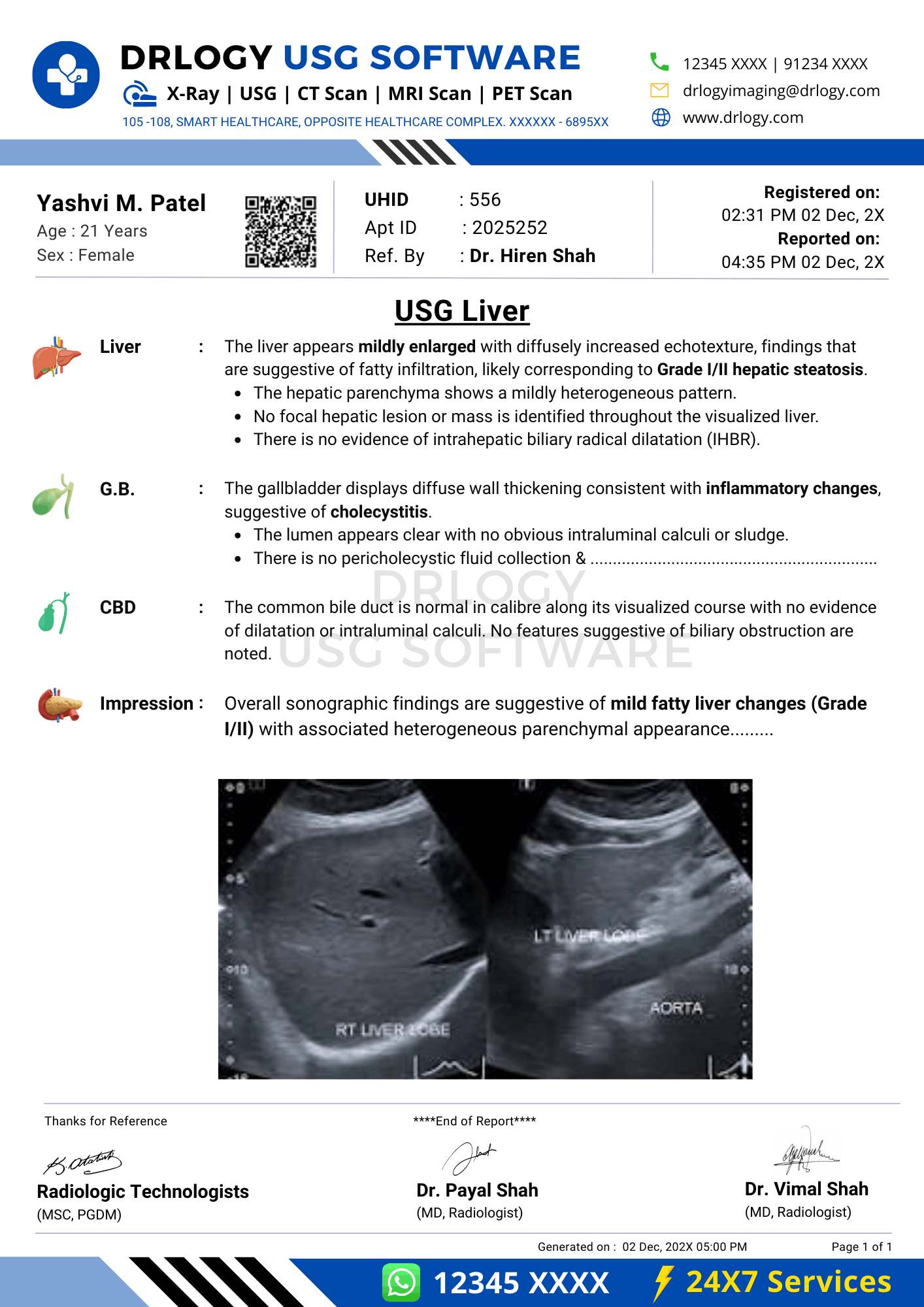
Normal Liver USG Report Format (Sample)
Patient & Study Information:
Patient: [Name], [Age]/[Sex]
Study Date/Time: [DD-MM-YYYY, Time]
Referrer: [Clinician/Department]
Examination: Liver Ultrasound
Clinical History / Indication:
Abnormal liver function tests. Evaluate liver parenchyma.
Technique / Protocol:
Transabdominal ultrasound of the liver performed using a curvilinear transducer. Standard subcostal and intercostal views obtained. Color Doppler assessment performed for portal vein and hepatic veins.
Findings:
Liver is normal in size with smooth surface contour. Parenchyma shows homogeneous echotexture with no focal lesionentified. No intrahepatic biliary dilatation is seen. Portal vein is normal in caliber with hepatopetal flow on Doppler. Hepatic veins appear patent. No perihepatic free fluid is noted.
Impression / Conclusion:
Normal sonographic appearance of the liver. No focal liver lesionentified.
Limitations:
No significant technical limitation noted.
Abnormal Liver USG Report Format (Sample)
Patient & Study Information:
Patient: [Name], [Age]/[Sex]
Study Date/Time: [DD-MM-YYYY, Time]
Referrer: [Clinician/Department]
Examination: Liver Ultrasound
Clinical History / Indication:
Known chronic liver disease. Surveillance evaluation.
Technique / Protocol:
Transabdominal ultrasound of the liver performed using a curvilinear transducer. Standard views obtained. Doppler assessment performed for portal venous flow.
Findings:
Liver appears mildly enlarged with altered echotexture and mildly irregular surface contour. A well-defined hyperechoic focal lesion is noted in the right lobe measuring approximately [x] x [y] x [z] cm; no definite internal vascularity is demonstrated on limited Doppler assessment. No intrahepatic biliary dilatation is seen. Portal vein caliber is mildly prominent with hepatopetal flow. Mild ascites is present.
Impression / Conclusion:
Sonographic features suggestive of diffuse parenchymal liver disease. Focal liver lesion as described; further characterization may be considered based on clinical context. Mild ascites noted.
Limitations:
Lesion characterization is limited on grayscale ultrasound; assessment partially limited by bowel gas/body habitus as applicable.
How Drlogy Radiology Reporting Software Standardizes These Report Formats
- Template-driven reporting supports consistent liver-specific sectioning (size, echotexture, surface, lesions, biliary/vascular assessment) across radiologists.
- Impression safety controls help maintain conservative medico-legal phrasing and reduce overstatement for indeterminate lesions.
- Uniform formatting across modalities enables consistent reporting across USG, CT, and MRI follow-up contexts within the same reporting ecosystem.
- AI-enabled reporting from scan images can assist in drafting structured text from standard checklists and repeated reporting patterns, while preserving radiologist verification and responsibility.
- Audit-ready documentation improves completeness checks, traceability of clinical notes, and structured data extraction for QA reviews.
Standardization features support quality and consistency without replacing clinical judgment.
10 Key Clinical Guidelines for an Effective Liver USG Report Format
- Document liver size using a consistent method and state whether it is normal, enlarged, or reduced in a reproducible manner.
- Describe surface contour explicitly (smooth vs irregular) and avoid etiologic conclusions unless supported by broader findings and context.
- Use standardized echogenicity/echeotexture terminology and include supportive descriptors such as beam attenuation when relevant.
- For focal lesions, record number, size in three dimensions, location (lobe/segment when feasible), margins, echogenicity, and posterior features.
- State presence or absence of intrahepatic biliary dilatation, and document porta hepatis observations when relevant to indication.
- Include portal vein caliber and flowection when Doppler is performed; document adequacy of Doppler assessment if limited.
- Document key ancillary findings that influence interpretation (ascites, perihepatic collections, visible splenic enlargement).
- Separate findings from impression; avoid repeating full findings in the impression and focus on clinically meaningful conclusions.
- Use conservative impression wording for indeterminate abnormalities and explicitly acknowledge limitations of ultrasound characterization.
- Include limitations transparently whenever acoustic window, body habitus, bowel gas, or preparation reduces sensitivity.
Consistent adherence to these guidelines improves diagnostic clarity, follow-up comparability, and medico-legal safety in daily liver ultrasound reporting.
Common Reporting Errors to Avoid
- Omitting portal vein assessment when Doppler is performed or when the indication suggests portal hypertension evaluation; prevent by using a mandatory vascular checklist.
- Reporting “fatty liver” or “cirrhosis” as definitive without describing supportive sonographic features; prevent by documenting objective features and using “suggestive of” wording.
- Incomplete lesion description (missing size, location, margins); prevent by structured lesion fields and standardized descriptors.
- Failure to document limitations (bowel gas, obesity, rib shadowing) despite reduced sensitivity; prevent by a fixed limitations subsection in every report.
- Overly broad impressions that do not address the referral question; prevent by aligning impression pointsectly to indication and key findings.
Avoiding these errors improves report credibility and clinical utility.
Medico-Legal Considerations in Radiology Reporting
- Documentation safety: record objective observations, measurements, and relevant negatives; avoid speculative statements not supported by images.
- Disclaimers: state study limitations clearly when sensitivity is reduced and avoid implying exclusion of disease beyond ultrasound capability.
- Accountability: ensure reportentifiers, signature/authorization, and study date/time are complete and traceable.
- Audit readiness: maintain consistent sectioning and terminology to support QA review, peer review, and structured audits.
- Comparative accuracy: when referencing prior studies, specify date/modality and use cautious interval language when imaging conditions differ.
- Conservative impressions: use differential-level wording for indeterminate lesions and avoid definitive oncologic labeling without adequate characterization.
- Record integrity: ensure final report version control and retention policies support medico-legal requirements and clinical continuity.
Medico-legal strength depends on completeness, conservative wording, and transparent documentation of scope and limitations.
Structured Reporting vs Narrative Reporting
| Aspect | Structured | Narrative |
|---|---|---|
| Completeness | Checklist-driven sections reduce omissions for size, lesions, biliary, and vascular items. | Dependent on individual memory and time; omissions occur more often in high-volume settings. |
| Consistency | Standard terminology and ordering improves multi-radiologist uniformity and follow-up comparability. | Style and terminology vary significantly across reporters and sites. |
| Audit readiness | Enables QA review, benchmarking, and structured data extraction for compliance. | Difficult to audit and benchmark; key elements may be non-searchable or inconsistently phrased. |
| Efficiency | Faster reporting for routine cases using templates with focused edits; reduces rework. | Can be fast for experienced users but often requires rework for missing items or unclear phrasing. |
| Medico-legal robustness | Clear scope, standardized limitations, and conservative impression framing improve defensibility. | Greater risk of ambiguous wording, missing limitations, and inconsistent documentation. |
Role of Technology in Radiology Reporting
- PACS and RIS integration supports seamless access to images, prior reports, and structured reporting workflows.
- Voice dictation improves efficiency when paired with structured templates that constrain section order and reduce omissions.
- AI-assisted formatting can standardize phrasing, populate templates, and prompt missing fields while preserving radiologist verification.
- RIS-based structured templates ensure uniform reporting across radiologists, shifts, and sites with controlled terminology.
- Modality-specific reporting software supports liver USG-specific checklists, Doppler fields, and lesion documentation consistency.
Technology improves consistency and throughput when it is designed to support, not replace, professional judgment.
Why High-Volume Radiology Centers Prefer Software-Based Reporting Formats
- Improved report turnaround time through templates and reduced rework for missing information.
- Higher quality assurance with standardized checklists for liver size, lesions, portal venous system, and biliary comments.
- Better multi-radiologist consistency across shifts and locations, supporting predictable clinical interpretation.
- Enhanced scalability as patient volumes rise, without proportional increase in variability or omission errors.
- Easier audit and compliance workflows through structured fields, report completeness scoring, and searchable terminology.
- Reduced medico-legal risk through consistent limitations documentation and conservative impression controls.
- Better longitudinal follow-up comparison by maintaining consistent structure and measurement reporting across serial studies.
Operational preference is driven by reliability, consistency, and audit readiness rather than stylistic preference alone.
Frequently Asked Questions (FAQs)
How should a Liver USG report format document echogenicity and echotexture without overstatement?
Use objective descriptors (homogeneous/heterogeneous, increased echogenicity, beam attenuation) and reserve etiologic statements for conservative impression language such as “suggestive of.”
What is the minimum lesion documentation required in a Liver USG report format?
Record lesion number, location, three-dimensional size, margins, echogenicity, internal architecture, posterior acoustic features, and Doppler vascularity description if assessed.
How should impressions be written for indeterminate focal liver lesions on ultrasound?
Summarize the lesion objectively and use non-definitive language; state that characterization is limited on grayscale ultrasound and recommend correlation or further evaluation if clinically indicated.
What medico-legal elements should always be present in a liver ultrasound report?
Clear patiententifiers, study date/time, technique scope, key positives and relevant negatives, documented limitations, and conservative impression wording aligned with ultrasound capability.
Does structured reporting reduce clinical flexibility compared with free-text dictation?
Structured reporting standardizes section order and completeness while allowing free-text within each section for nuanced interpretation and clinically appropriate emphasis.
Key Takeaways for Radiology Professionals
- Use a consistent liver USG report structure to reduce omissions in size, echotexture, lesion description, and portal/biliary documentation.
- Document objective findings with standardized terminology and place interpretation conservatively in the impression.
- For focal lesions, ensure reproducible lesion characterization: location, size in three dimensions, margins, echogenicity, internal features, and Doppler pattern when available.
- Include vascular comments (portal vein caliber/flow) when clinically relevant and document Doppler limitations explicitly when present.
- Treat limitations as mandatory documentation whenever sensitivity is reduced by bowel gas, body habitus, rib shadowing, or preparation issues.
- Align the impression with the referral question and avoid definitive diagnostic labeling when ultrasound characterization is inherently limited.
- Maintain audit-ready documentation through structured templates, controlled terminology, and consistent section ordering across radiologists and sites.
Expert Picks
Final Conclusion
A standardized Liver USG report format is essential for consistent clinical communication, reliable follow-up comparison, and medico-legal safety in professional radiology practice.
Structured reporting software, when aligned with real-world radiologist workflows, supports completeness and consistency while maintaining the radiologist’s interpretive authority and accountability, consistent with Drlogy’s reporting approach.