Drlogy
Healthcare organization
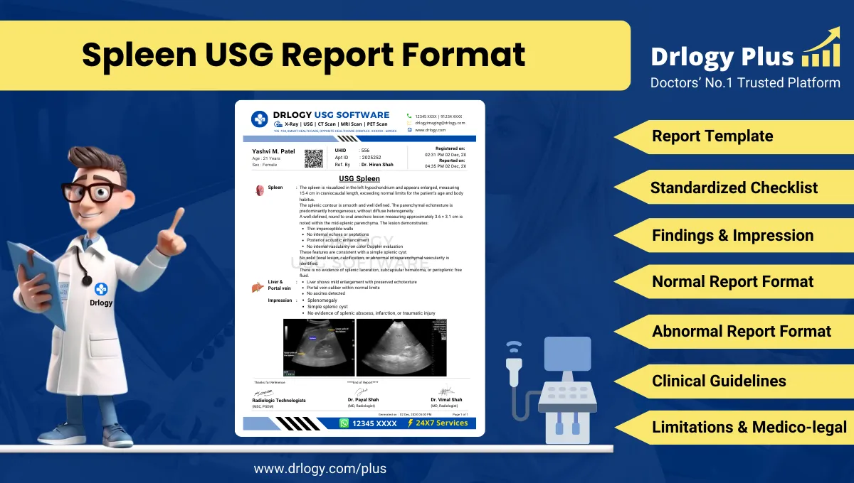
Spleen USG Report Format for Radiologists
What Is a Spleen USG Report Format?
A Spleen USG report format is a standardized professional structure for documenting ultrasonographic evaluation of the spleen using consistent terminology and measurement methodology.
It functions as a clinical communication tool supporting diagnosis, referral decisions, follow-up assessment, and longitudinal comparison across serial imaging studies.
It is also a medico-legal document defining examination scope, technical adequacy, objective findings, conservative interpretation, and stated limitations in accordance with accepted radiology standards.
Check:
Explore the Best AI-Based Ultrasound Reporting Software for Radiologists
Clinical Importance of a Standardized Spleen USG Report Format
- Diagnostic clarity by ensuring systematic documentation of splenic size, echotexture, focal lesions, vascular status, and perisplenic findings.
- Inter-doctor communication through consistent terminology for splenomegaly grading, focal splenic lesions, infarcts, and traumatic changes.
- Reporting consistency across radiologists and centers, particularly for follow-up of splenomegaly, portal hypertension, and hematological disorders.
- Patient safety by reducing omission of critical elements such as splenic length, volume estimation context, and perisplenic fluid assessment.
- Medico-legal protection by documenting technique, limitations, and conservative impression language aligned with ultrasound capabilities.
A standardized report format improves reliability, reproducibility, and audit readiness in routine and specialized splenic ultrasound reporting.
Why Manual Reporting Often Fails to Maintain Standardization at Scale
- Inter-radiologist variability in describing splenic size, echogenicity, and lesion characteristics leads to inconsistent interpretation.
- Missed sections in high-volume settings commonly include splenic measurements, perisplenic space evaluation, or comparison statements.
- Terminology inconsistency such as borderline splenomegaly versus mild enlargement complicates follow-up and clinical decision-making.
- Audit challenges arise because narrative free-text reports lack structured data points required for quality assurance and benchmarking.
Software-assisted structured reporting improves completeness and uniformity without replacing radiologist expertise.
Indications for Spleen USG
- Evaluation of suspected splenomegaly
- Follow-up of known splenic enlargement in hematological or hepatic disorders
- Assessment in portal hypertension and chronic liver disease
- Evaluation of focal splenic lesions detected on prior imaging
- Post-trauma assessment for splenic injury in stable patients
- Fever of unknown origin with suspected splenic involvement
- Suspected splenic infarction or abscess as clinically indicated
- Monitoring splenic size in hematological malignancies or infections
A focused indication ensures clinically relevant reporting and a targeted impression.
Pre-Examination Details to Be Documented
- Patiententifiers including name, age, sex, unique, accession number, study date and time.
- Referral details including referring clinician, department, and clinical indication.
- Clinical notes such as history of liver disease, hematological disorders, trauma, infection, or prior splenic imaging.
- Preparation status including fasting status when relevant and factors affecting acoustic window.
- Safety checks including patient verification and correct study labeling.
How Reporting Software Ensures Complete Pre-Examination Documentation
- Mandatory field enforcement ensuresentifiers, indication, and clinical context are documented before report finalization.
- Safety checklist compliance standardizes patient verification and study confirmation.
- Clinical note traceability links referral details and prior imaging contextectly to the report.
- Implementation example: Drlogy Radiology Reporting Software supports structured splenic ultrasound templates with mandatory pre-examination fields and standardized phrasing controls.
Standard Sections of a Spleen USG Report Format
- Patient & Study Information
- Clinical History / Indication
- Technique / Protocol
- Findings (splenic evaluation and related structures)
- Impression / Conclusion
- Limitations of the Study
- Recommendations & Follow-Up (if applicable)
A consistent section order improves readability, completeness, and medico-legal defensibility.
Patient & Study Information Section
This section establishes traceability and accountability:
- Patient demographics and uniqueentifiers
- Study date, time, and accession number
- Referring clinician and department
- Examination name and scope
- Prior imaging references if available
Clinical History / Indication Section
Best practices include:
- Concise documentation of the clinical question
- Inclusion of relevant history such as chronic liver disease, hematological disorders, trauma, or infection
- Avoidance of speculative or unrelated information
Technique / Protocol Section
- Positioning: supine and right lateral decubitus as required for optimal splenic visualization.
- Views: longitudinal and transverse splenic views; subcostal and intercostal approaches as needed.
- Transducer: curvilinear probe appropriate for depth; linear probe for superficial splenic lesions when required.
- Doppler usage: documented when splenic or portal vascular assessment is performed.
- Measurements: splenic length measured in the longest axis; additional dimensions recorded per institutional practice.
Technique documentation defines examination scope and supports interpretation reliability.
Findings Section – Organ/System-Wise Reporting
Splenic findings should be documented objectively and systematically.
Spleen
- Visualization: adequately visualized or partially obscured; note factors affecting assessment.
- Size: splenic length measured in centimeters; categorized conservatively as normal or enlarged, with grading if used institutionally.
- Echotexture: homogeneous or heterogeneous; relative echogenicity described objectively.
- Margins and contour: smooth or irregular.
- Focal lesions:
Cystic lesions described by size, wall, internal echoes, septations, and posterior enhancement.
- Solid lesions described by size, echogenicity, margins, and Doppler vascularity if assessed.
- Splenic hilum and vessels: assessed when indicated; document Doppler findings conservatively.
- Perisplenic region: presence or absence of free fluid, collection, or hematoma when applicable.
Associated Findings
- Liver and portal system: brief contextual comment if relevant to splenic findings, avoiding duplication of full hepatic reporting.
- Ascites: documented if present.
- Trauma-related findings: describe laceration, hematoma, or irregular echotexture conservatively without injury grading unless institutional protocol allows.
Objective description should precede interpretation, with explicit documentation of normal findings when present.
Impression / Conclusion Section
- Summarize key findings relevant to the indication.
- Use conservative, non-definitive language such as “features suggestive of.”
- Avoid etiological certainty where ultrasound sensitivity is limited.
- Include relevant negatives when clinically important.
- Recommend correlation with clinical and laboratory findings when appropriate.
Limitations of the Study
- Bowel gas limiting splenic visualization
- Patient body habitus reducing acoustic penetration
- Limited intercostal window
- Suboptimal breath-hold or patient cooperation
- Limited characterization of indeterminate focal lesions on ultrasound
Documenting limitations ensures transparency and medico-legal safety.
Recommendations & Follow-Up (If Applicable)
- Correlate splenic size and echotexture with clinical and laboratory findings.
- Consider follow-up ultrasound for interval assessment when clinically indicated.
- Consider cross-sectional imaging for further characterization of indeterminate splenic lesions when appropriate.
Recommendations must remain conservative and indication-linked.
Normal Spleen USG Report Format (Sample)
Patient & Study Information:
Patient: [Name], [Age]/[Sex]
Study Date: [DD-MM-YYYY]
Examination: Spleen Ultrasound
Clinical History / Indication:
Evaluate splenic size.
Technique / Protocol:
Ultrasound examination of the spleen performed using a curvilinear transducer with intercostal approach.
Findings:
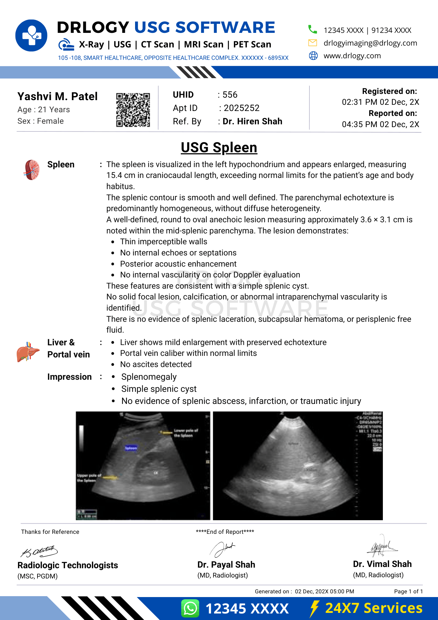
The spleen is normal in size measuring [ ] cm in length. Echotexture is homogeneous with smooth margins. No focal splenic lesion isentified. No perisplenic free fluid is seen.
Impression / Conclusion:
Normal sonographic appearance of the spleen.
Limitations:
No significant technical limitation noted.
Abnormal Spleen USG Report Format (Sample)
Patient & Study Information:
Patient: [Name], [Age]/[Sex]
Study Date: [DD-MM-YYYY]
Examination: Spleen Ultrasound
Clinical History / Indication:
Chronic liver disease. Evaluate splenic size.
Technique / Protocol:
Ultrasound examination of the spleen performed using standard intercostal views.
Findings:
The spleen is enlarged measuring [ ] cm in length. Echotexture appears mildly heterogeneous. No focal splenic lesion isentified. Mild ascites is noted in the perisplenic region.
Impression / Conclusion:
Splenomegaly as described. Clinical correlation is advised.
Limitations:
Assessment limited by suboptimal intercostal acoustic window.
How Drlogy Radiology Reporting Software Standardizes These Report Formats
- Template-driven reporting ensures consistent splenic size measurement and lesion documentation.
- Impression safety controls promote conservative wording and avoid diagnostic overstatement.
- Uniform formatting across modalities supports continuity between USG and cross-sectional imaging.
- AI-enabled reporting assistance supports structured content generation under radiologist verification.
- Audit-ready documentation facilitates quality assurance and medico-legal review.
10 Key Clinical Guidelines for an Effective Spleen USG Report Format
- Always document splenic length using a consistent measurement technique.
- Use conservative terminology for splenomegaly grading.
- Describe echotexture objectively before interpretation.
- Explicitly document absence of focal lesions when none are seen.
- Assess and document perisplenic region when clinically relevant.
- Use Doppler findings cautiously and document only when performed.
- Avoid etiological conclusions unsupported by ultrasound findings.
- Separate findings from impression clearly.
- Document limitations whenever sensitivity is reduced.
- Maintain consistent report structure across examinations.
Adhering to these guidelines improves diagnostic reliability and medico-legal safety.
Common Reporting Errors to Avoid
- Omission of splenic measurements
- Inconsistent splenomegaly terminology
- Overinterpretation of heterogeneous echotexture
- Missing documentation of perisplenic fluid
- Failure to state technical limitations
Avoidance of these errors improves report credibility.
Medico-Legal Considerations in Radiology Reporting
- Objective documentation of findings
- Explicit limitation statements
- Conservative impression language
- Clear accountability and report authorization
- Audit-ready structure
- Appropriate use of disclaimers
- Accurate comparison statements
Medico-legal robustness depends on completeness and conservative interpretation.
Structured Reporting vs Narrative Reporting
| Aspect | Structured | Narrative |
|---|---|---|
| Completeness | Standardized fields | Variable |
| Consistency | High | Operator dependent |
| Audit readiness | Strong | Limited |
| Efficiency | Optimized | Variable |
| Medico-legal safety | Enhanced | Variable |
Role of Technology in Radiology Reporting
- PACS and RIS integration
- Voice dictation with templates
- AI-assisted formatting
- RIS-based structured templates
- Modality-specific reporting tools
Technology enhances consistency without replacing professional judgment.
Why High-Volume Radiology Centers Prefer Software-Based Reporting Formats
- Faster turnaround time
- Improved quality assurance
- Multi-radiologist consistency
- Better scalability
- Reduced omission errors
- Enhanced audit readiness
- Improved medico-legal safety
Frequently Asked Questions (FAQs)
What defines a standard Spleen USG report format?
A structured format documenting size, echotexture, focal lesions, impression, and limitations using conservative terminology.
How should splenomegaly be reported?
By objective measurement and conservative grading without etiological assumption.
Are focal splenic lesions diagnosable on ultrasound?
Ultrasound can describe morphology; definitive diagnosis often requires correlation or further imaging.
Why are limitations important in spleen ultrasound reporting?
They document reduced sensitivity and protect medico-legal integrity.
Key Takeaways for Radiology Professionals
- A standardized Spleen USG report format ensures consistent documentation of size, echotexture, and focal lesions.
- Conservative language, explicit limitations, and structured templates improve clinical communication, follow-up comparability, and medico-legal safety.
Expert Picks
Final Conclusion
A standardized Spleen USG report format is essential for accurate clinical interpretation, longitudinal assessment, and medico-legal defensibility in routine and specialized imaging practice.
Structured reporting software supports completeness, consistency, and conservative impression formulation while aligning with real-world radiology workflows and professional standards.