Drlogy
Healthcare organization
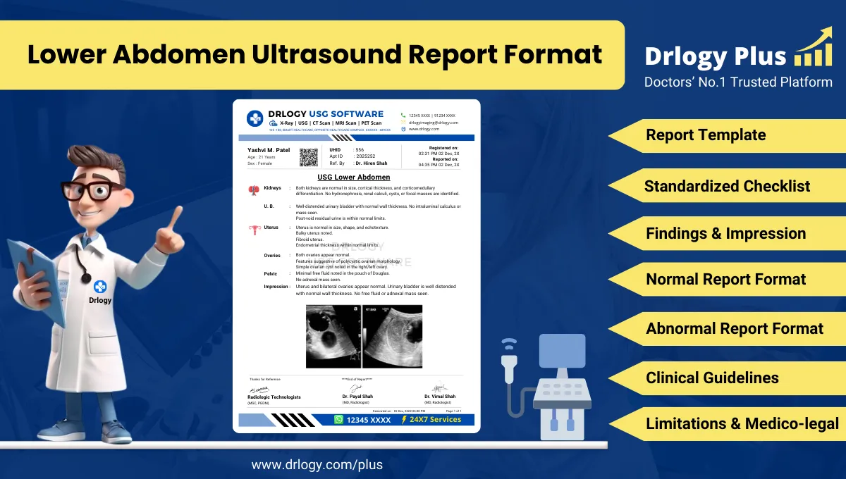
Lower Abdomen Ultrasound Report Format for Radiologists
What Is a Lower Abdomen Ultrasound Report Format?
A Lower abdomen ultrasound report format is a standardized professional framework for documenting ultrasonographic evaluation of pelvic and lower abdominal organs with clarity and consistency.
Clinically, it ensures accurate communication of findings relevant to urogenital, gynecological, and pelvic pathology across diagnostic, referral, and follow-up workflows.
Medico-legally, it functions as a formal record defining examination scope, technical adequacy, interpretation, and limitations in accordance with accepted reporting standards.
Check:
Explore the Best AI-Based Ultrasound Reporting Software for Radiologists
Clinical Importance of a Standardized Lower Abdomen Ultrasound Report Format
Lower abdominal ultrasound examinations frequently involve pelvic organs, urinary structures, and adjacent soft tissues, where interpretation is highly dependent on systematic documentation.
Structured reporting improves professional practice in the following ways:
- Diagnostic clarity
Organ-wise structured documentation reduces ambiguity when evaluating pelvic organs, urinary bladder, uterus, adnexa, prostate, and adjacent spaces. - Inter-doctor communication
Standardized terminology and section flow improve comprehension between radiologists, gynecologists, urologists, surgeons, and referring physicians. - Reporting consistency
Uniform formats minimize subjective variation across radiologists, technicians, and reporting environments, especially in high-volume centers. - Patient safety
Complete documentation decreases the likelihood of missed pelvic findings, incomplete evaluation, or misinterpretation during follow-up. - Medico-legal protection
Clear documentation of findings, impressions, and limitations demonstrates adherence to professional standards during audits and legal review.
Why Manual Reporting Often Fails to Maintain Standardization at Scale
Manual or free-text reporting often becomes inconsistent when applied across large patient volumes and multiple reporting doctors.
Professionally observed limitations include:
- Significant inter-radiologist variability in terminology, structure, and emphasis
- Missed or inadequately described pelvic structures during time pressure
- Inconsistent measurement reporting and follow-up comparability
- Difficulty in internal audits, peer review, and quality benchmarking
Software-assisted structured reporting mitigates these risks by enforcing consistent sectioning and completeness without constraining clinical interpretation.
Indications for Lower Abdomen Ultrasound
Common, clinically relevant indications include:
- Pelvic pain or lower abdominal pain
- Evaluation of urinary bladder pathology
- Assessment of uterus and adnexa
- Prostate evaluation
- Suspected pelvic mass
- Abnormal menstrual history or gynecologic symptoms
- Urinary retention or lower urinary tract symptoms
- Follow-up of known pelvic pathology
Pre-Examination Details to Be Documented
Accurate pre-examination documentation is essential for interpretation accuracy and medico-legal safety.
Mandatory elements include:
- Patient name, age, sex, and uniqueentification number
- Referring clinician, department, and clinical specialty
- Clear clinical indication and relevant history
- Preparation status, including bladder filling
- Basic safety and procedural verification
Incomplete pre-examination data may compromise diagnostic reliability.
How Reporting Software Ensures Complete Pre-Examination Documentation
Structured reporting systems support documentation integrity through:
- Mandatory patiententifier fields
- Enforced clinical indication entry
- Preparation checklist validation
- Traceable linkage between referral notes and imaging findings
Drlogy Radiology Reporting Software may be cited as an implementation example where such controls are integrated into routine radiology workflows.
Standard Sections of a Lower Abdomen Ultrasound Report Format
A universally accepted lower abdomen ultrasound report format includes:
- Patient and Study Information
- Clinical History / Indication
- Technique / Protocol
- Findings (organ-wise)
- Impression / Conclusion
- Limitations of the Study
- Recommendations and Follow-Up, if applicable
Patient & Study Information Section
This section establishes traceability and must include:
- Patient demographics
- Study date and time
- Accession or study number
- Referring clinician details
Accurateentifiers are essential for longitudinal comparison and audit readiness.
Clinical History / Indication Section
Best practices include:
- Concise, relevant documentation focused on pelvic or urinary symptoms
- Inclusion of relevant laboratory findings or prior imaging
- Avoidance of unrelated or speculative clinical information
This section contextualizes imaging findings and supports clinical correlation.
Technique / Protocol Section
For lower abdomen ultrasound, documentation should include:
- Patient positioning (supine, oblique if required)
- Transducer type and frequency
- Transabdominal approach details
- Bladder filling adequacy
- Use of additional focused views, if performed
The technique section defines the technical scope and inherent limitations of the study.
Findings Section – Organ/System-Wise Reporting
The findings section is the diagnostic core of the report and must be objective and systematic.
Best practices include:
- Describing findings before interpretation
- Explicitly documenting normal structures
- Reporting abnormalities with size, echotexture, margins, and location
- Maintaining consistent terminology
Commonly evaluated structures include:
- Urinary bladder
- Uterus (if applicable)
- Endometrium
- Ovaries and adnexa
- Prostate (if applicable)
- Seminal vesicles
- Pelvic free fluid
- Adjacent soft tissues
Impression / Conclusion Section
The impression should:
- Summarize key findings concisely
- Use conservative, non-definitive language
- Include differential considerations where appropriate
- Avoid clinical overstatement beyond imaging findings
This section guides clinical decision-making while maintaining medico-legal safety.
Limitations of the Study
Limitations must be documented whenever present.
Common examples include:
- Inadequate bladder distension
- Bowel gas interference
- Patient body habitus
- Limited visualization of deep pelvic structures
Documenting limitations protects diagnostic integrity.
Recommendations & Follow-Up (If Applicable)
Recommendations should be:
- Clinically appropriate
- Conservative and non-directive
- Clearly separated from diagnostic conclusions
Normal Lower Abdomen Ultrasound Report Format (Sample)
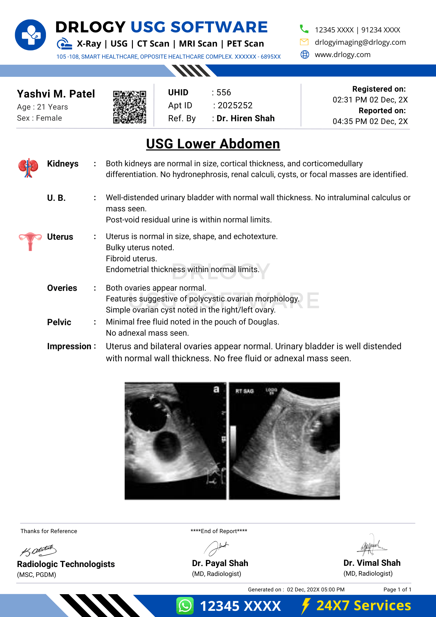
Findings:
The urinary bladder is adequately distended with normal wall thickness. No intraluminal mass or calculus is seen. Uterus appears normal in size and echotexture. Endometrial thickness is within normal limits. Both ovaries are normal in size and echotexture. No adnexal mass or pelvic free fluid is noted.
Impression:
Normal ultrasonography of the lower abdomen.
Abnormal Lower Abdomen Ultrasound Report Format (Sample)
Findings:
The urinary bladder shows mild wall thickening. Uterus is mildly enlarged with heterogeneous echotexture. A well-defined cystic lesion is noted in the right adnexa. Mild pelvic free fluid is present.
Impression:
Ultrasonographic findings as described above. Clinical correlation is advised.
How Drlogy Radiology Reporting Software Standardizes These Report Formats
Clinically relevant standardization mechanisms include:
- Template-driven organ-wise reporting
- Impression safety controls to prevent overstatement
- Uniform formatting across ultrasound and other modalities
- AI-enabled draft generation from imaging workflows
- Audit-ready structured documentation
No pricing, promotional language, or call-to-action elements are included in clinical reporting.
10 Key Clinical Guidelines for an Effective Lower Abdomen Ultrasound Report Format
- Maintain consistent section order
- Document all evaluated pelvic organs
- Explicitly state normal findings
- Use standardized anatomical terminology
- Avoid speculative diagnostic language
- Separate findings from impressions
- Document limitations clearly
- Verify patiententifiers
- Keep impressions concise
- Support longitudinal comparison
Adherence to these principles improves quality and medico-legal safety.
Common Reporting Errors to Avoid
- Omission of pelvic structures
- Over-interpretation of equivocal findings
- Inconsistent terminology
- Failure to document limitations
- Non-standard impression phrasing
Medico-Legal Considerations in Radiology Reporting
Key medico-legal considerations include:
- Complete and accurate documentation
- Conservative diagnostic language
- Clear accountability of the reporting radiologist
- Explicit documentation of limitations
- Audit and peer-review readiness
- Secure record retention
- Consistency with accepted reporting standards
Structured Reporting vs Narrative Reporting
| Aspect | Structured Reporting | Narrative Reporting |
|---|---|---|
| Consistency | High | Variable |
| Audit readiness | Strong | Limited |
| Efficiency | Optimized | Operator dependent |
Role of Technology in Radiology Reporting
Technology supports radiology reporting through:
- PACS and RIS integration
- Voice dictation with structured templates
- AI-assisted formatting and consistency checks
- RIS-based structured reporting modules
- Modality-specific reporting software
Why High-Volume Radiology Centers Prefer Software-Based Reporting Formats
Operational advantages include:
- Reduced report turnaround time
- Improved quality assurance
- Consistency across multiple radiologists
- Scalable reporting workflows
- Enhanced audit readiness
- Reduced reporting errors
- Uniform documentation across modalities
Frequently Asked Questions (FAQs)
What is the standard structure of a lower abdomen ultrasound report format?
A structured sequence including patient details, technique, findings, impression, and limitations.
Should normal pelvic organs be explicitly documented?
Yes. Explicit documentation improves clarity, follow-up comparison, and medico-legal safety.
How detailed should the impression be?
Concise, summary-focused, and limited to imaging findings.
Is structured reporting mandatory for pelvic ultrasound?
Not mandatory, but strongly recommended in professional radiology practice.
Does software-based reporting replace radiologist judgment?
No. Software enhances consistency while preserving clinical expertise.
Key Takeaways for Radiology Professionals
- Standardized formats improve diagnostic reliability
- Structured reporting reduces omissions and variability
- Conservative impressions protect professional integrity
- Technology enhances, not replaces, clinical judgment
Expert Picks
Final Conclusion
A standardized Lower abdomen ultrasound report format is essential for accurate clinical communication, patient safety, and medico-legal protection in modern radiology practice.
Structured reporting systems, when aligned with real-world workflows, enable consistency in high-volume environments while fully respecting the radiologist’s professional judgment and responsibility.