Drlogy
Healthcare organization
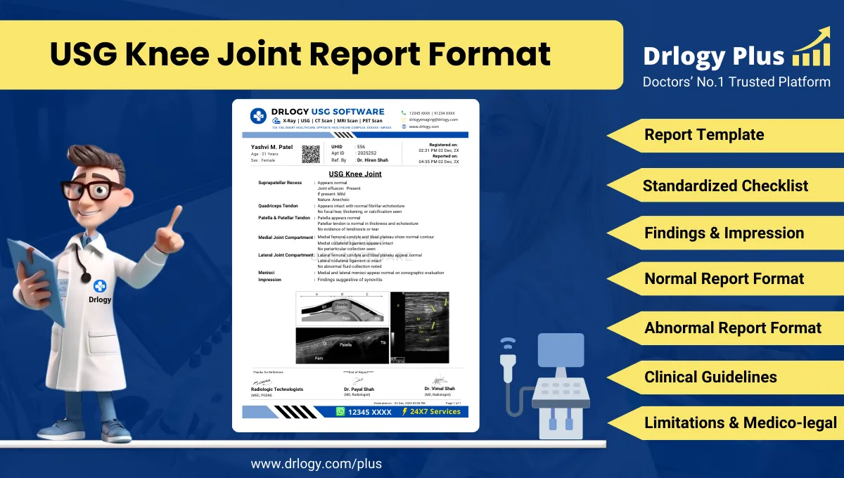
USG Knee Joint Report Format for Radiologists
What Is a USG Knee Joint Report Format?
A USG knee joint report format is a standardized professional structure for documenting ultrasonographic evaluation of the knee joint and periarticular soft tissues.
It serves as a clinical communication record supporting diagnosis, referrals, follow-up, and comparison with prior musculoskeletal imaging.
It is a medico-legal document defining examination scope, technical adequacy, objective findings, conservative interpretation, and documented limitations.
Check:
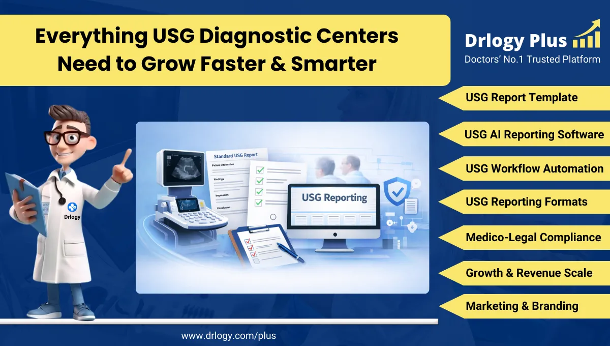
Best AI-Based Ultrasound Reporting Software for Radiologists
Clinical Importance of a Standardized USG Knee Joint Report Format
- Diagnostic clarity by systematically documenting tendons, ligaments, joint recesses, bursae, and periarticular soft tissues.
- Inter-doctor communication through uniform terminology understood by radiologists, orthopedicians, sports physicians, and rheumatologists.
- Reporting consistency across serial examinations, different operators, and high-volume musculoskeletal practices.
- Patient safety by minimizing omission of key structures such as quadriceps tendon, patellar tendon, suprapatellar recess, and medial collateral ligament.
- Medico-legal protection through objective description, standardized impressions, and explicit limitation statements.
A standardized report format improves reliability and longitudinal comparability in knee ultrasound reporting.
Why Manual Reporting Often Fails to Maintain Standardization at Scale
- Inter-radiologist variability in describing effusions, synovial hypertrophy, tendon abnormalities, and ligament appearances.
- Missed sections in high-volume settings such as incomplete documentation of recesses or collateral ligaments.
- Terminology inconsistency including vague descriptors without anatomical localization.
- Audit challenges because narrative reports lack structured musculoskeletal parameters.
Software-assisted structured reporting improves completeness and medico-legal robustness.
Indications for USG Knee Joint
- Knee pain evaluation
- Suspected joint effusion or synovitis
- Soft tissue swelling around the knee
- Tendon or ligament injury assessment
- Follow-up of inflammatory or degenerative conditions
- Guidance for aspiration or injection
A focused indication guides protocol selection and reporting emphasis.
Pre-Examination Details to Be Documented
- Patiententifiers including name, age, sex, unique, accession number, study date, and time.
- Referral details including referring clinician and clinical query.
- Clinical notes including pain location, duration, trauma history, and prior imaging if provided.
- Preparation status including patient positioning and ability to cooperate.
- Safety checks including correct patient verification and side confirmation.
How Reporting Software Ensures Complete Pre-Examination Documentation
- Mandatory field enforcement for indication, laterality, and examination scope.
- Safety checklist compliance ensuring correct patient and sideentification.
- Clinical note traceability linking referral information with imaging findings.
- Implementation example: Drlogy Radiology Reporting Software provides structured musculoskeletal templates with compulsory knee joint assessment fields.
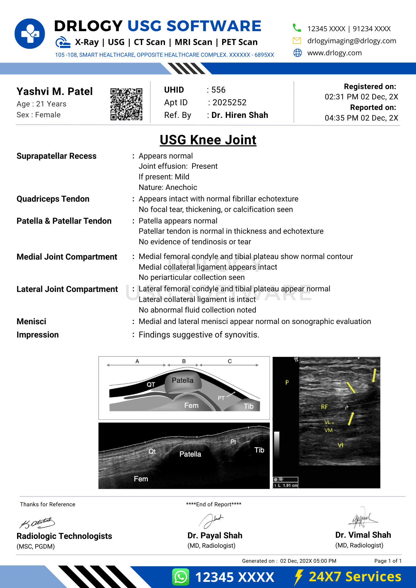
Standard Sections of a USG Knee Joint Report Format
- Patient & Study Information
- Clinical History / Indication
- Technique / Protocol
- Findings (structure-wise knee assessment)
- Impression / Conclusion
- Limitations of the Study
- Recommendations & Follow-Up (if applicable)
Patient & Study Information Section
This section ensures traceability and accountability:
- Patient demographics andentifiers
- Study date, time, and accession number
- Referring clinician details
- Examination name and laterality
- Comparison with prior imaging if available
Clinical History / Indication Section
- Presenting symptoms and duration
- Side involved
- History of trauma or inflammation
- Relevant prior interventions or imaging
Documentation must remain concise and clinically relevant.
Technique / Protocol Section
- Positioning: supine with knee in slight flexion.
- Approach: high-frequency linear transducer examination.
- Views: anterior, medial, lateral, and posterior knee compartments.
- Dynamic assessment: real-time evaluation during flexion or stress where indicated.
- Doppler usage: documented when assessing synovial vascularity.
Technique documentation defines examination adequacy and interpretive confidence.
Findings Section – Organ/System-Wise Reporting
Suprapatellar Recess
- Presence or absence of joint effusion
- Fluid characteristics
- Synovial thickening if present
Quadriceps Tendon
- Thickness and echotexture
- Focal defects or calcifications
Patellar Tendon
- Continuity and thickness
- Enthesopathy features
Medial and Lateral Collateral Ligaments
- Fiber continuity
- Thickening or focal hypoechoic areas
Meniscal Periphery (Accessible Portions)
- Extrusion or parameniscal cysts when visualized
Posterior Knee
- Baker’s cyst assessment
- Popliteal fossa soft tissues
Objective description must clearly separate normal from abnormal findings.
Impression / Conclusion Section
- Concise summary of key abnormalities
- Conservative, non-definitive language
- Avoidance of etiological or prognostic overstatement
- Correlation with clinical findings advised
Limitations of the Study
- Limited visualization of intra-articular structures
- Operator dependency
- Patient discomfort or restricted movement
- Acoustic shadowing from bone
Limitations must be explicitly documented for medico-legal clarity.
Recommendations & Follow-Up (If Applicable)
- Clinical correlation with symptoms
- Follow-up imaging when indicated
- MRI correlation if deeper intra-articular evaluation is required
Recommendations must remain conservative and appropriate.
Normal USG Knee Joint Report Format (Sample)
- Patient & Study Information:
- Patient: [Name], [Age]
- Study Date: [DD-MM-YYYY]
Examination: USG Knee Joint
Clinical History / Indication:
Knee pain.
Technique / Protocol:
Ultrasound examination of the knee performed using high-frequency transducer.
Findings:
No joint effusion is seen. Quadriceps and patellar tendons appear intact. Collateral ligaments show normal echotexture. No focal soft tissue abnormality isentified.
Impression / Conclusion:
No significant sonographic abnormality detected in the knee joint.
Limitations:
No significant technical limitation noted.
Abnormal USG Knee Joint Report Format (Sample)
- Patient & Study Information
- Patient: [Name], [Age]
- Study Date: [DD-MM-YYYY]
Examination: USG Knee Joint
Clinical History / Indication:
Swelling around knee.
Technique / Protocol:
Ultrasound examination performed.
Findings:
A moderate suprapatellar joint effusion is noted. Mild synovial thickening is present. Quadriceps and patellar tendons are intact.
Impression / Conclusion:
Sonographic findings as described. Clinical correlation is advised.
Limitations:
Assessment limited by patient discomfort.
How Drlogy Radiology Reporting Software Standardizes These Report Formats
- Template-driven reporting ensuring systematic knee structure documentation
- Impression safety controls enforcing conservative wording
- Uniform formatting across musculoskeletal studies
- AI-enabled reporting assistance under radiologist supervision
- Audit-ready documentation supporting quality assurance
10 Key Clinical Guidelines for an Effective USG Knee Joint Report Format
- Document laterality clearly.
- Assess all accessible knee compartments.
- Describe effusion objectively.
- Evaluate tendons and ligaments systematically.
- Use standardized anatomical terminology.
- Separate findings from impression.
- Avoid definitive intra-articular diagnoses.
- Document limitations explicitly.
- Compare with prior studies when available.
- Maintain consistent report structure.
Adherence improves reporting accuracy and medico-legal safety.
Common Reporting Errors to Avoid
- Omission of suprapatellar recess assessment
- Vague tendon descriptions
- Failure to document laterality
- Overinterpretation of ultrasound findings
- Missing limitation statements
Avoidance of these errors strengthens report reliability.
Medico-Legal Considerations in Radiology Reporting
- Objective documentation of findings
- Standardized terminology usage
- Conservative impression language
- Explicit limitation statements
- Clear accountability
- Audit-ready structure
- Appropriate disclaimers
Structured Reporting vs Narrative Reporting
| Aspect | Structured | Narrative |
|---|---|---|
| Completeness | High | Variable |
| Consistency | Standardized | Operator dependent |
| Audit readiness | Strong | Limited |
| Efficiency | Optimized | Variable |
| Legal safety | Enhanced | Variable |
Role of Technology in Radiology Reporting
- PACS and RIS integration
- Voice dictation
- AI-assisted formatting
- RIS-based structured templates
- Musculoskeletal workflow tools
Technology enhances consistency without replacing professional judgment.
Why High-Volume Radiology Centers Prefer Software-Based Reporting Formats
- Faster turnaround time
- Improved quality assurance
- Multi-radiologist consistency
- Scalability across MSK studies
- Reduced omission errors
- Standardized documentation
- Enhanced medico-legal protection
Frequently Asked Questions (FAQs)
What defines a standard USG knee joint report format?
A structured format documenting periarticular soft tissues, impression, and limitations using standardized musculoskeletal terminology.
Can ultrasound evaluate all knee structures?
Ultrasound evaluates accessible soft tissues and must be correlated with MRI for intra-articular structures.
How should effusions be reported?
By objective description of presence, extent, and characteristics.
Why are limitations essential in knee ultrasound reports?
They define modality constraints and support medico-legal defensibility.
Key Takeaways for Radiology Professionals
- Use standardized structure for every knee ultrasound.
- Document all accessible compartments systematically.
- Maintain conservative impression wording.
- Explicitly state technical limitations.
Consistent structured reporting improves musculoskeletal ultrasound reliability.
Expert Picks
Final Conclusion
A standardized USG knee joint report format is essential for accurate musculoskeletal imaging communication, reliable follow-up, and medico-legal safety in clinical practice.
Structured reporting software supports consistency, completeness, and conservative interpretation while aligning with real-world radiology workflows and professional standards.