Drlogy
Healthcare organization
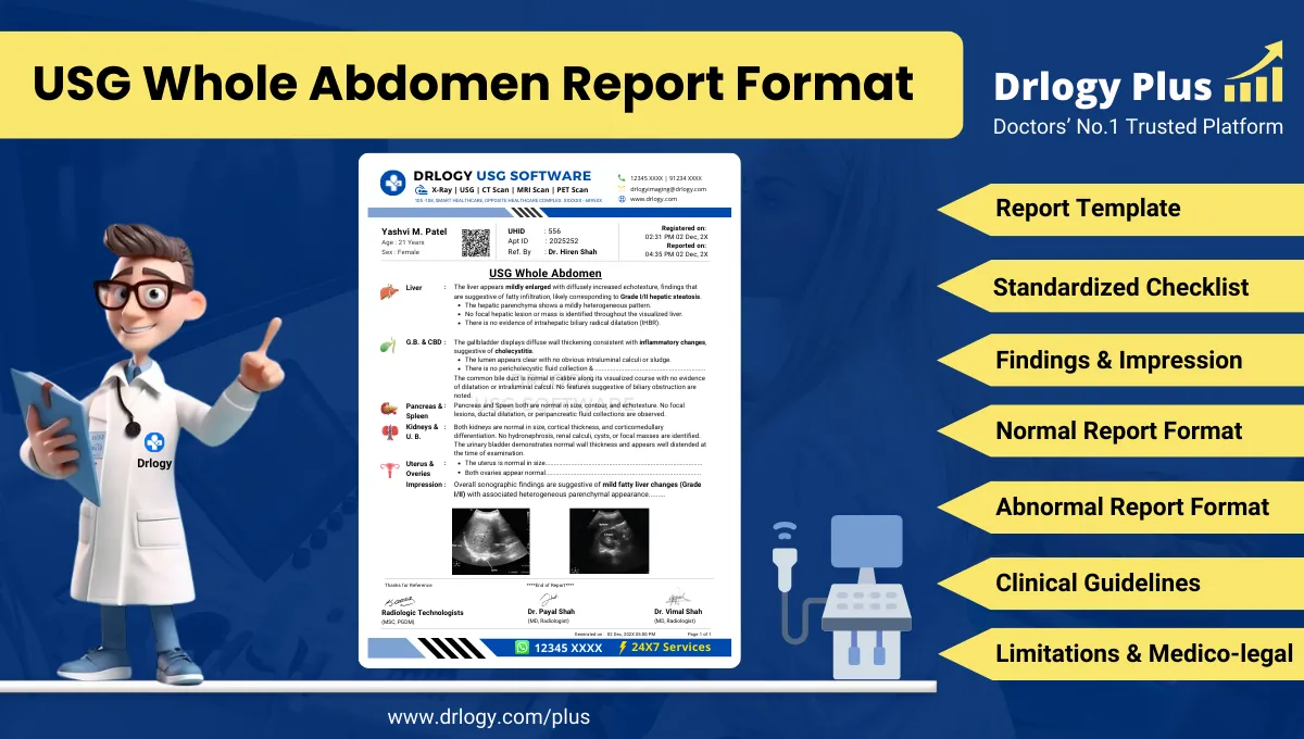
USG Whole Abdomen Report Format for Radiologists
What Is a USG Whole Abdomen Report Format?
A USG whole abdomen report format is a standardized professional structure for documenting comprehensive ultrasonographic evaluation of all abdominal organs in a single examination.
It ensures systematic documentation of findings to support diagnosis, referrals, follow-up decisions, and longitudinal comparison across serial imaging studies.
It serves as a medico-legal record defining examination scope, technique, observations, interpretation, and documented limitations in accordance with accepted radiology standards.
Check:
Explore the Best AI-Based Ultrasound Reporting Software for Radiologists
Clinical Importance of a Standardized USG Whole Abdomen Report Format
Structured reporting improves professional practice through the following:
- Diagnostic clarity by ensuring complete organ-wise assessment
- Improved inter-doctor communication across specialties
- Consistency of reporting in multi-radiologist environments
- Enhanced patient safety through reduced omission errors
- Strong medico-legal protection via standardized documentation
Why Manual Reporting Often Fails to Maintain Standardization at Scale
Manual narrative reporting commonly faces limitations in high-volume diagnostic settings.
Inter-radiologist variability leads to inconsistent structure and terminology, even for similar findings. Under time pressure, sections may be incompletely documented or missed entirely.
Terminology inconsistency complicates clinical interpretation and follow-up comparison. Free-text reports are also difficult to audit, standardize, and benchmark during quality assurance and medico-legal review.
Software-assisted structured reporting addresses these challenges by enforcing completeness and consistency while preserving radiologist judgment.
Indications for USG Whole Abdomen
Common clinical indications include:
- Acute or chronic abdominal pain
- Evaluation of hepatobiliary pathology
- Renal and urinary tract assessment
- Suspected abdominal mass or organomegaly
- Evaluation of ascites
- Abnormal liver or renal function tests
- Follow-up of known abdominal disease
Pre-Examination Details to Be Documented
Essential pre-examination documentation includes:
- Patient name, age, sex, and uniqueentifier
- Referring clinician and department
- Clear clinical indication and relevant history
- Preparation status such as fasting
- Basic safety and procedural verification
Accurate documentation at this stage is critical for interpretation accuracy and medico-legal defensibility.
How Reporting Software Ensures Complete Pre-Examination Documentation
Structured reporting systems ensure completeness through:
- Mandatory patiententifier fields
- Required clinical indication entry
- Preparation and safety checklist compliance
- Traceable linkage between referral information and imaging findings
Drlogy Radiology Reporting Software is an example of an implementation where such safeguards are integrated into routine reporting workflows.
Standard Sections of a USG Whole Abdomen Report Format
Universally accepted sections include:
- Patient and Study Information
- Clinical History / Indication
- Technique / Protocol
- Findings (organ-wise)
- Impression / Conclusion
- Limitations of the Study
- Recommendations and Follow-Up, if applicable
Patient & Study Information Section
This section must document:
- Patient demographics
- Study date and time
- Accession or study number
- Referring clinician details
Theseentifiers ensure traceability, audit readiness, and longitudinal comparison.
Clinical History / Indication Section
Best practices include:
- Concise documentation focused on the clinical question
- Inclusion of relevant laboratory results or prior imaging
- Avoidance of unrelated or speculative information
Technique / Protocol Section
For USG whole abdomen, documentation should include:
- Patient positioning
- Transducer type and frequency
- Real-time scanning approach
- Use of Doppler evaluation where applicable
The technique section defines the technical scope and limitations of the examination.
Findings Section – Organ/System-Wise Reporting
The findings section is the diagnostic core of the report and must be objective and systematic.
Best practices include:
- Description before interpretation
- Explicit documentation of normal organs
- Abnormal findings described with size, echotexture, margins, and location
- Consistent anatomical terminology
Commonly evaluated organs include:
- Liver
- Gallbladder and biliary tree
- Pancreas
- Spleen
- Kidneys
- Urinary bladder
- Major abdominal vessels
- Peritoneal cavity
Impression / Conclusion Section
The impression should:
- Summarize key findings concisely
- Use conservative, non-definitive language
- Include differential considerations where appropriate
- Avoid diagnostic overstatement
Limitations of the Study
Limitations must be documented when present, such as:
- Bowel gas obscuration
- Patient body habitus
- Suboptimal acoustic window
- Incomplete visualization of specific organs
Recommendations & Follow-Up (If Applicable)
Recommendations should be:
- Clinically appropriate
- Conservative and non-directive
- Clearly separated from diagnostic conclusions
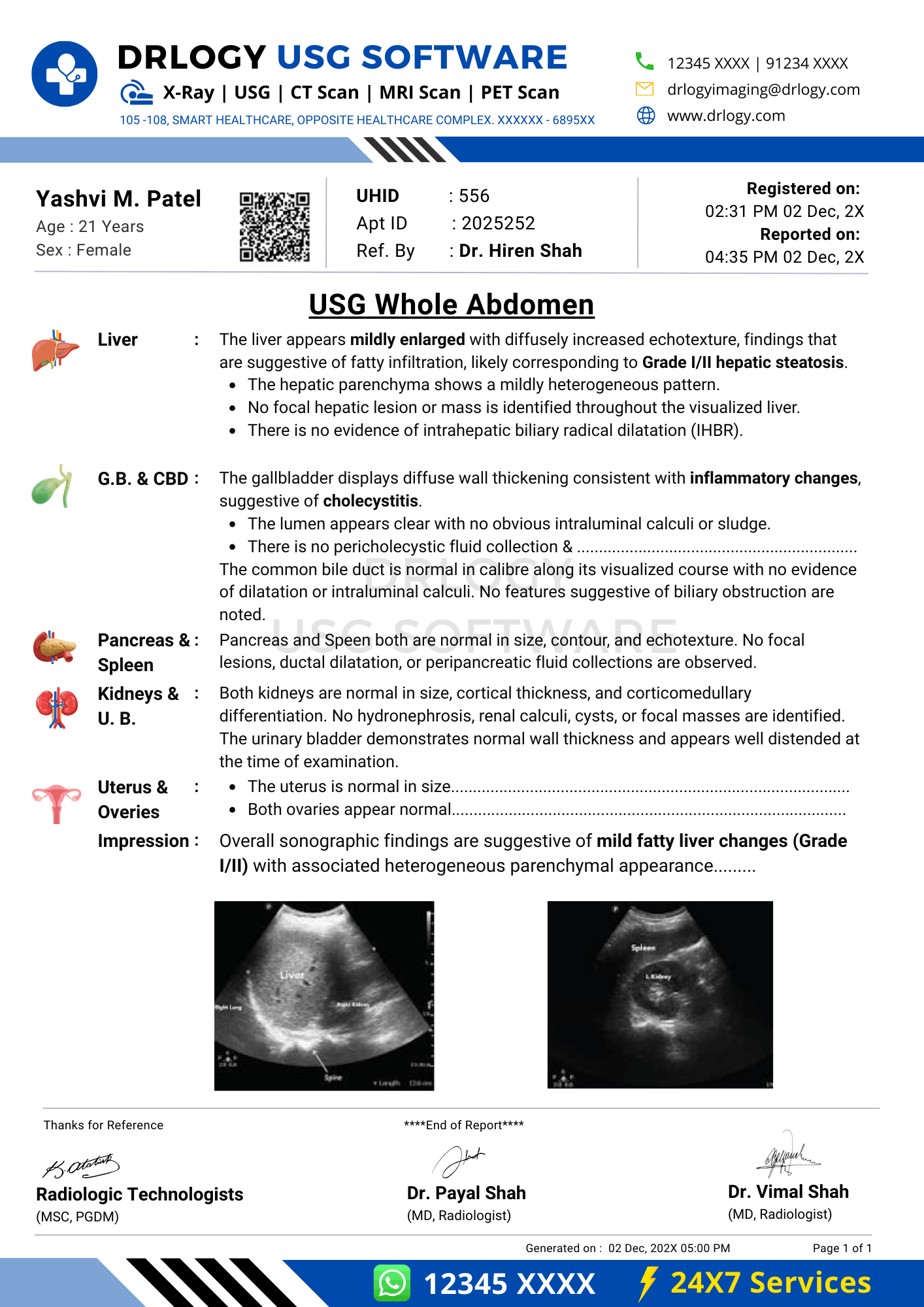
Normal USG Whole Abdomen Report Format (Sample)
Findings:
The liver is normal in size with homogeneous echotexture. No focal lesionentified. Gallbladder is well distended with normal wall thickness. No calculus or sludge noted. Common bile duct is within normal limits. Pancreas and spleen appear normal. Both kidneys are normal in size and echotexture with preserved corticomedullary differentiation. Urinary bladder is adequately distended with normal wall thickness. No free fluid is seen.
Impression:
Normal ultrasonography of the whole abdomen.
Abnormal USG Whole Abdomen Report Format (Sample)
Findings:
The liver is mildly enlarged with altered echotexture. Gallbladder shows an echogenic focus with posterior acoustic shadowing. Mild splenomegaly is noted. Both kidneys show normal size with mild pelvicalyceal dilatation on the right side. Minimal free fluid is present in the abdomen.
Impression:
Ultrasonographic findings as described above. Clinical correlation is advised.
How Drlogy Radiology Reporting Software Standardizes These Report Formats
Clinically relevant standardization features include:
- Template-driven organ-wise reporting
- Impression safety controls to prevent overstatement
- Uniform formatting across imaging modalities
- AI-enabled reporting assistance from imaging workflows
- Audit-ready structured documentation
10 Key Clinical Guidelines for an Effective USG Whole Abdomen Report Format
- Maintain consistent section order
- Document all evaluated organs
- Explicitly state normal findings
- Use standardized anatomical terminology
- Avoid speculative language
- Separate findings from impressions
- Document limitations clearly
- Verify patiententifiers
- Keep impressions concise
- Support longitudinal comparison
Following these guidelines improves diagnostic accuracy and medico-legal safety.
Common Reporting Errors to Avoid
- Omission of organ documentation
- Over-interpretation of equivocal findings
- Inconsistent terminology
- Failure to document limitations
- Non-standard impression phrasing
Medico-Legal Considerations in Radiology Reporting
Key medico-legal principles include:
- Complete and accurate documentation
- Conservative diagnostic language
- Clear accountability of the reporting radiologist
- Documentation of study limitations
- Audit and peer-review readiness
- Secure record retention
- Consistency with accepted reporting standards
Structured Reporting vs Narrative Reporting
| Aspect | Structured Reporting | Narrative Reporting |
|---|---|---|
| Consistency | High | Variable |
| Audit readiness | Strong | Limited |
| Efficiency | Optimized | Operator dependent |
Role of Technology in Radiology Reporting
Technology supports reporting through:
- PACS and RIS integration
- Voice dictation combined with structured templates
- AI-assisted formatting and consistency checks
- RIS-based structured reporting modules
- Modality-specific reporting software
Why High-Volume Radiology Centers Prefer Software-Based Reporting Formats
Operational benefits include:
- Reduced report turnaround time
- Improved quality assurance
- Consistency across multiple radiologists
- Scalability of reporting workflows
- Enhanced audit readiness
- Reduced reporting errors
- Uniform documentation across modalities
Frequently Asked Questions (FAQs)
What is the standard structure of a USG whole abdomen report format?
A structured sequence including patient details, technique, findings, impression, and limitations.
Should normal abdominal organs be explicitly documented?
Yes. Explicit documentation improves clarity and medico-legal safety.
How detailed should the impression be?
Concise and limited strictly to imaging findings.
Is structured reporting mandatory for whole abdomen ultrasound?
Not mandatory, but strongly recommended in professional practice.
Key Takeaways for Radiology Professionals
- Standardized formats improve diagnostic reliability
- Structured reporting reduces omission and variability
- Conservative impressions protect professional integrity
- Technology enhances, not replaces, clinical judgment
Expert Picks
Final Conclusion
A standardized usg whole abdomen report format is essential for accurate clinical communication, patient safety, and medico-legal protection in modern radiology practice.
Structured reporting systems aligned with real-world workflows support consistency, audit readiness, and professional accountability while fully respecting radiologist expertise and responsibility.